Patient Preparation and Positioning
Examinations should be scheduled in the morning.
Patients are asked to remain NPO overnight and take 2 tablets of simethicone the night before and the morning of the examination to reduce bowel gas.
Patients are asked to empty their urinary bladder immediately before the examination.
Patients are asked to remove any restrictive undergarments and informed that a drape will be placed over them for privacy.
Patients are placed in the supine position with the head of the bed elevated 10-15 degrees.
The examination starts with the anterior approach.
If excessive bowel gas is observed, the transducer is used to massage the bowel gas out of view. Alternatively, the patient is placed into the right or left lateral decubitus position.
The linear transducer is utilized for the infra-inguinal iliac system.
The curvilinear transducer is utilized for the supra-inguinal iliac system and inferior vena cava.
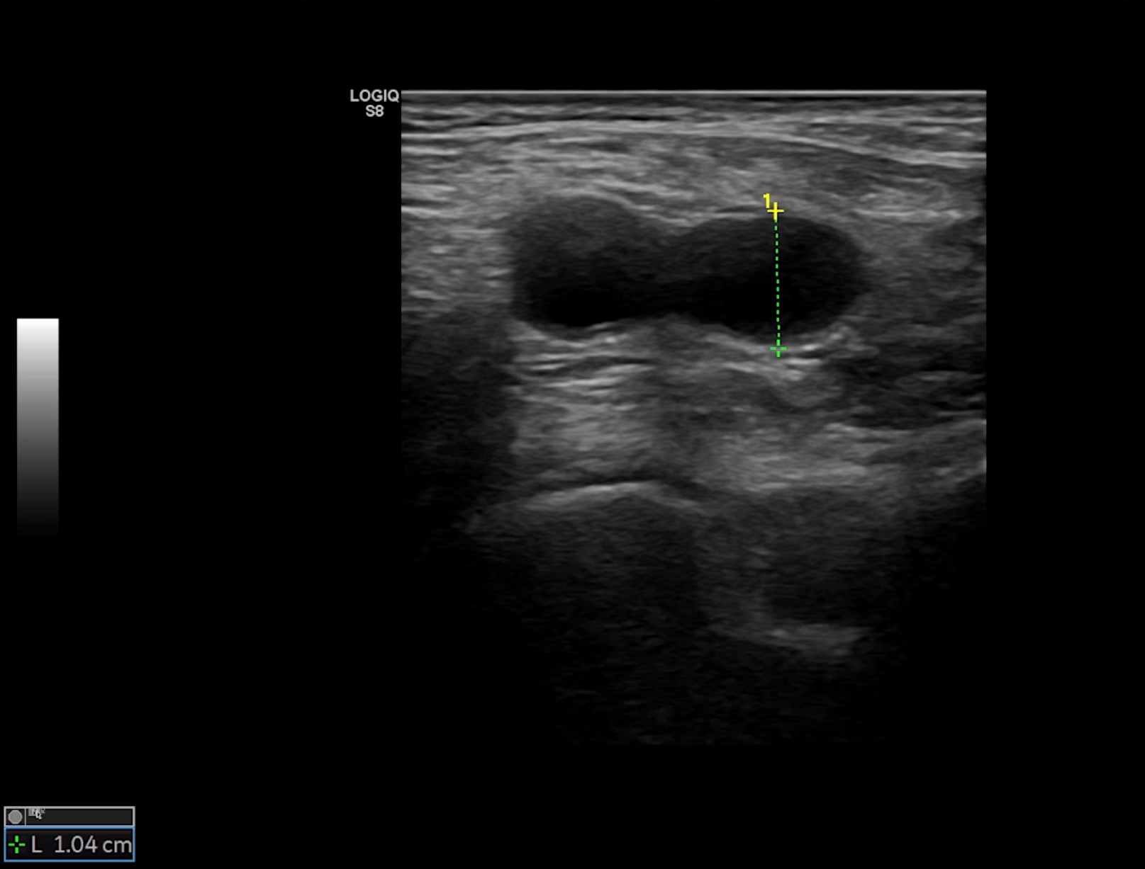
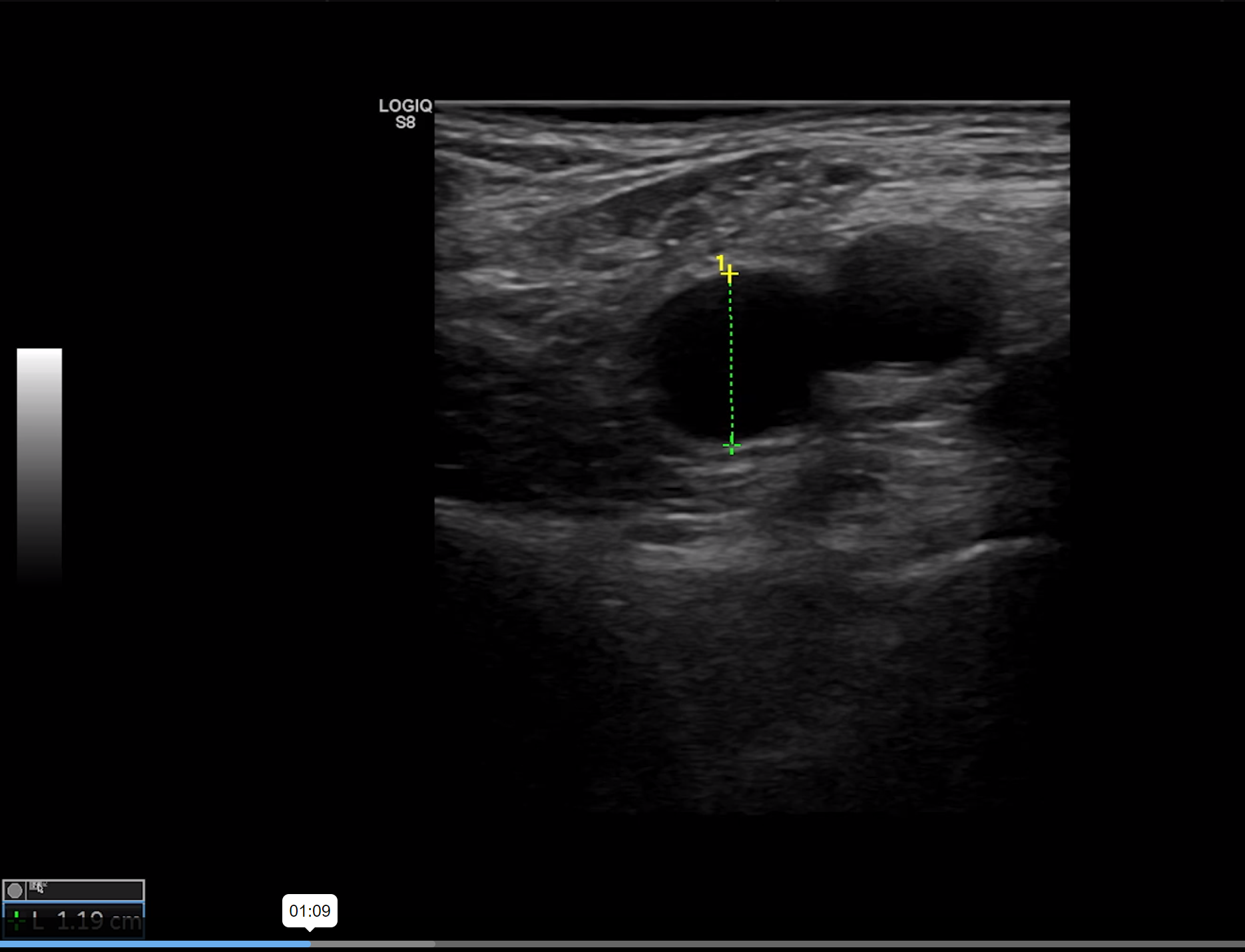
1. Right Common Femoral Vein:
B-mode, diameter measurements from anterior to posterior wall and medial to lateral wall at the largest diameter
Transverse View
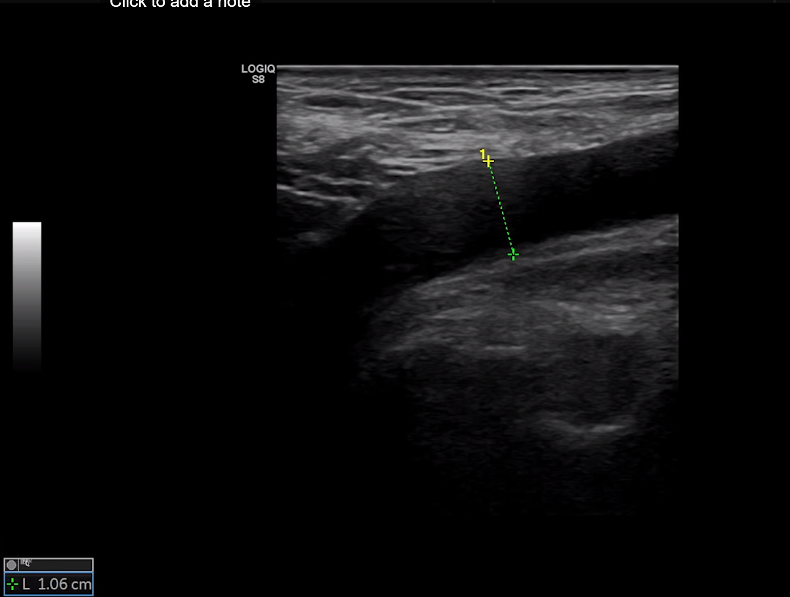
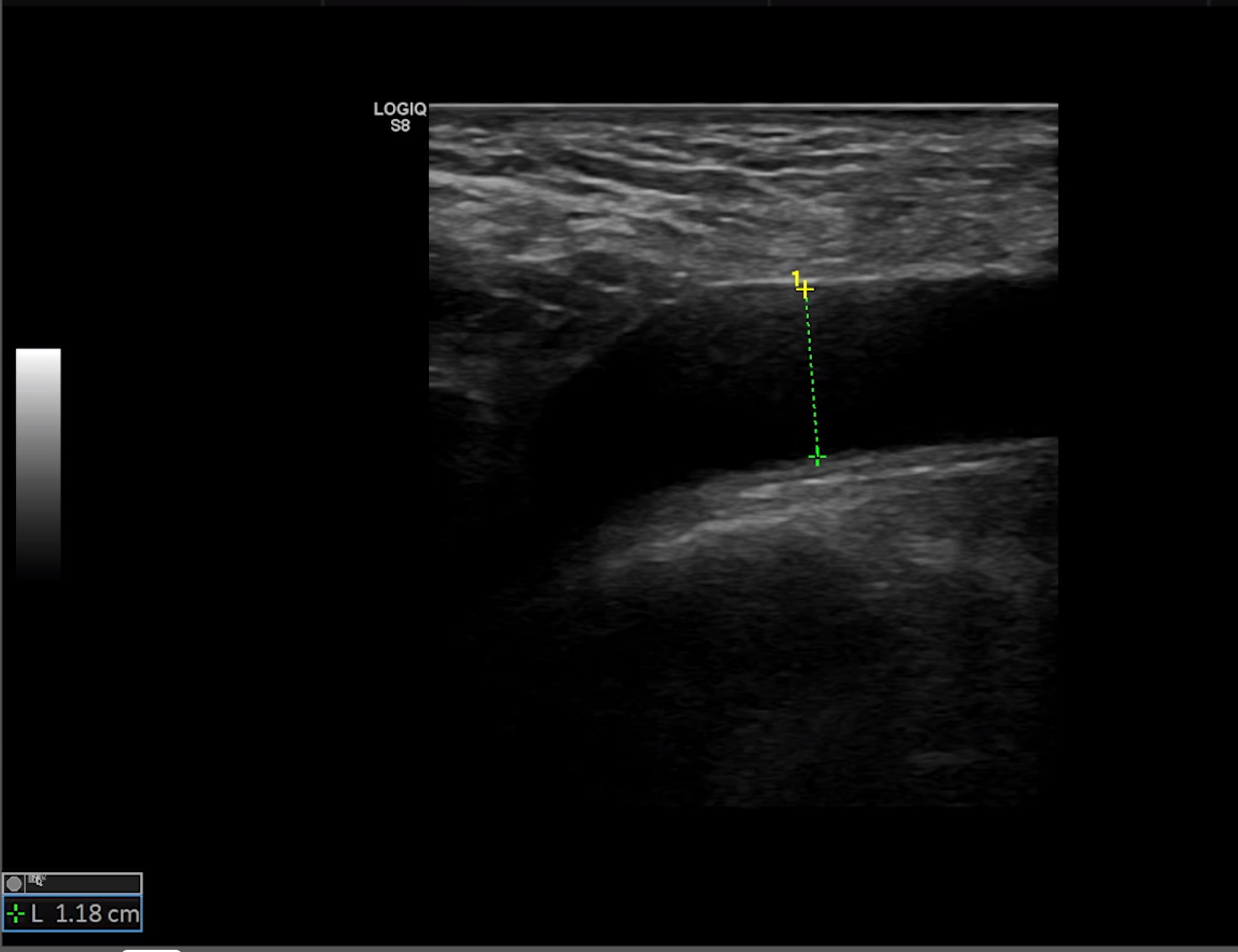
2. Right Common Femoral Vein:
B-mode, diameter measurements from anterior to posterior wall at the largest diameter
Longitudinal View
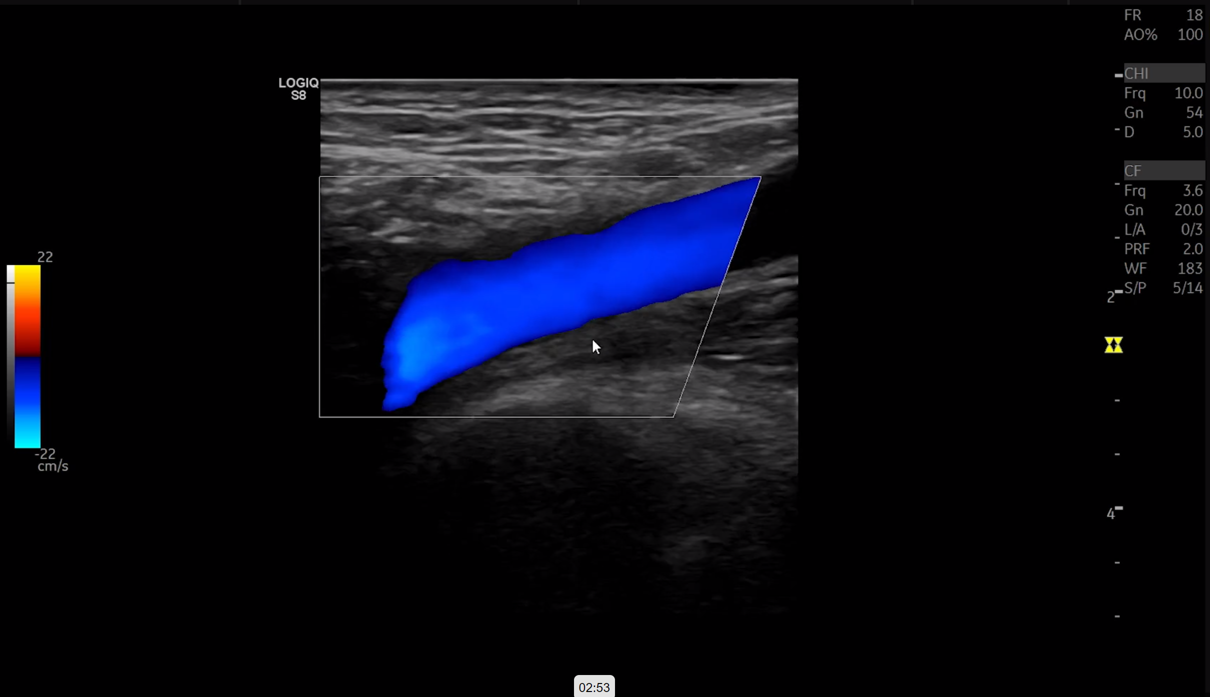
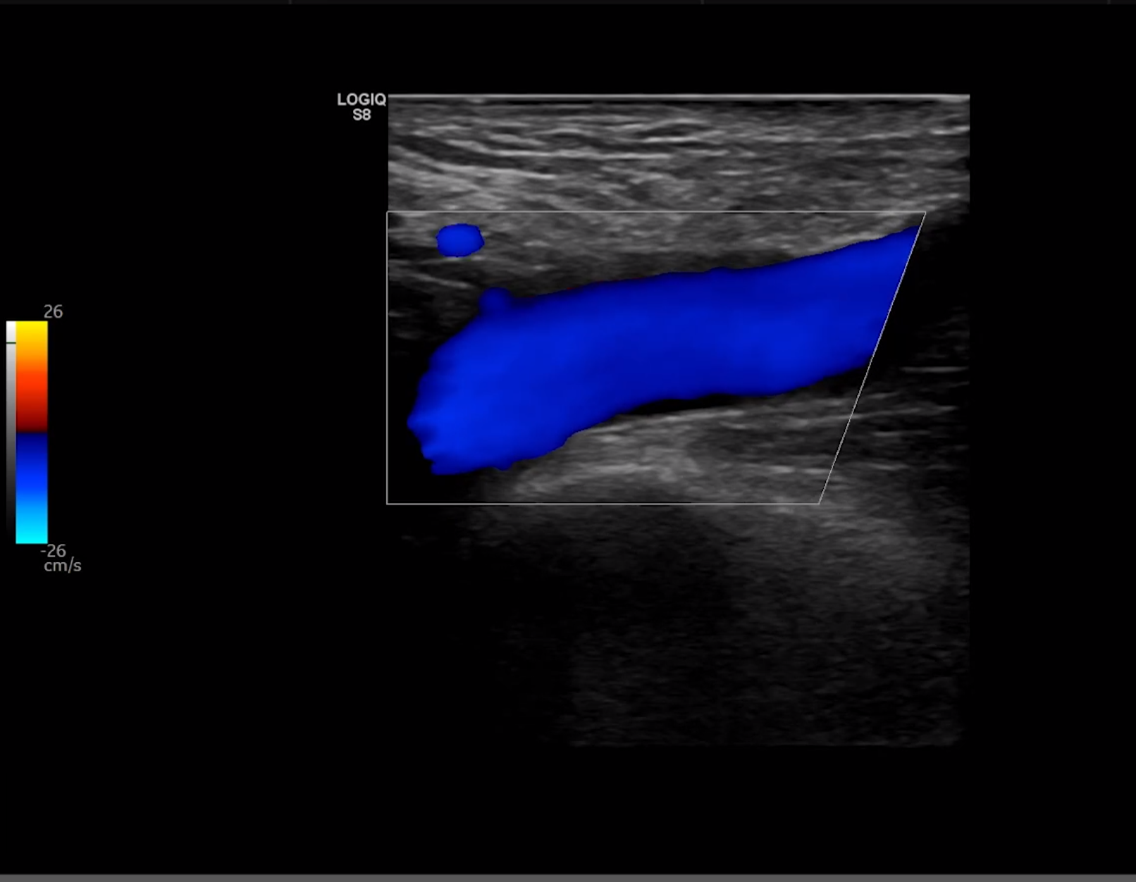
3. Right Common Femoral Vein:
Color Doppler
Longitudinal View
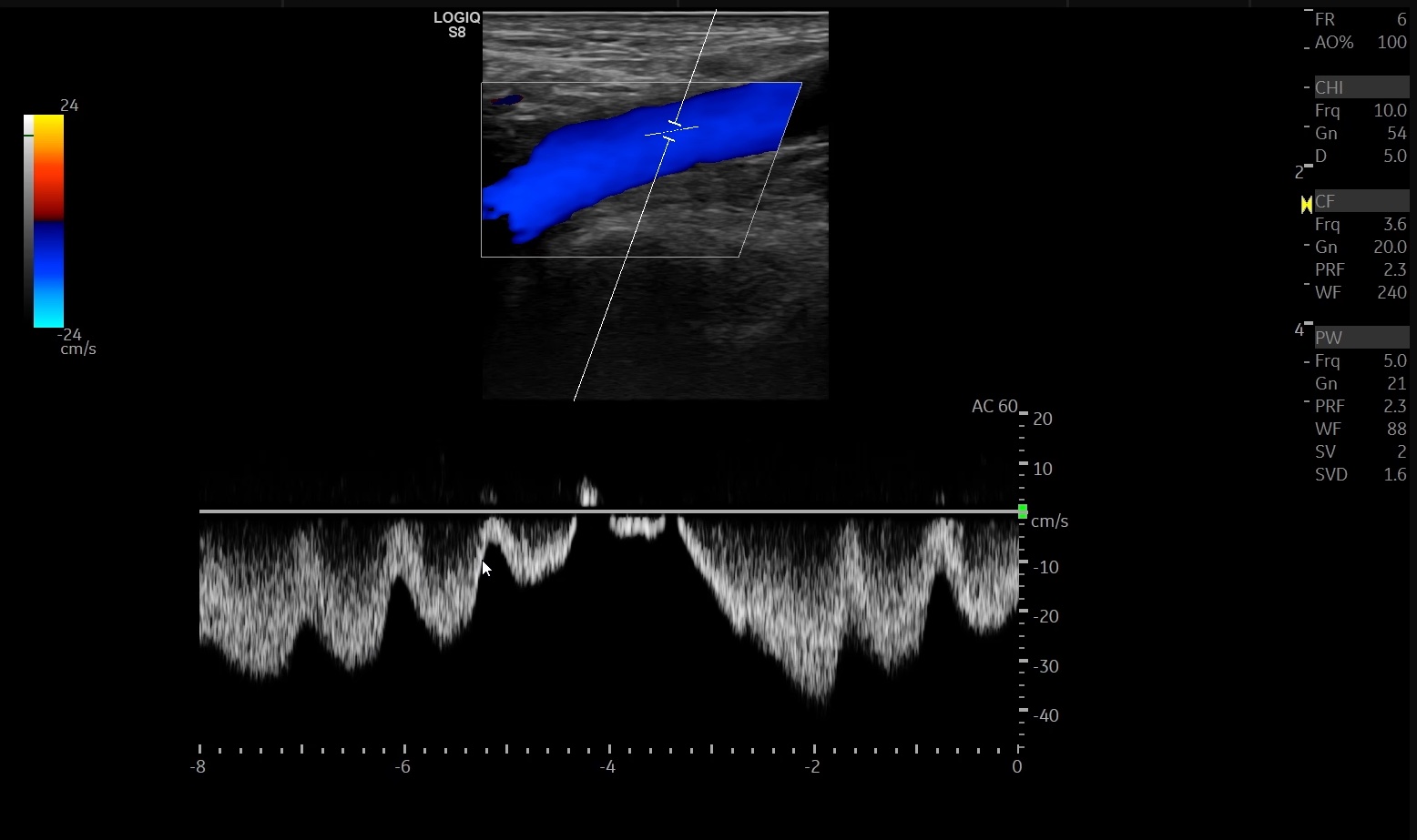
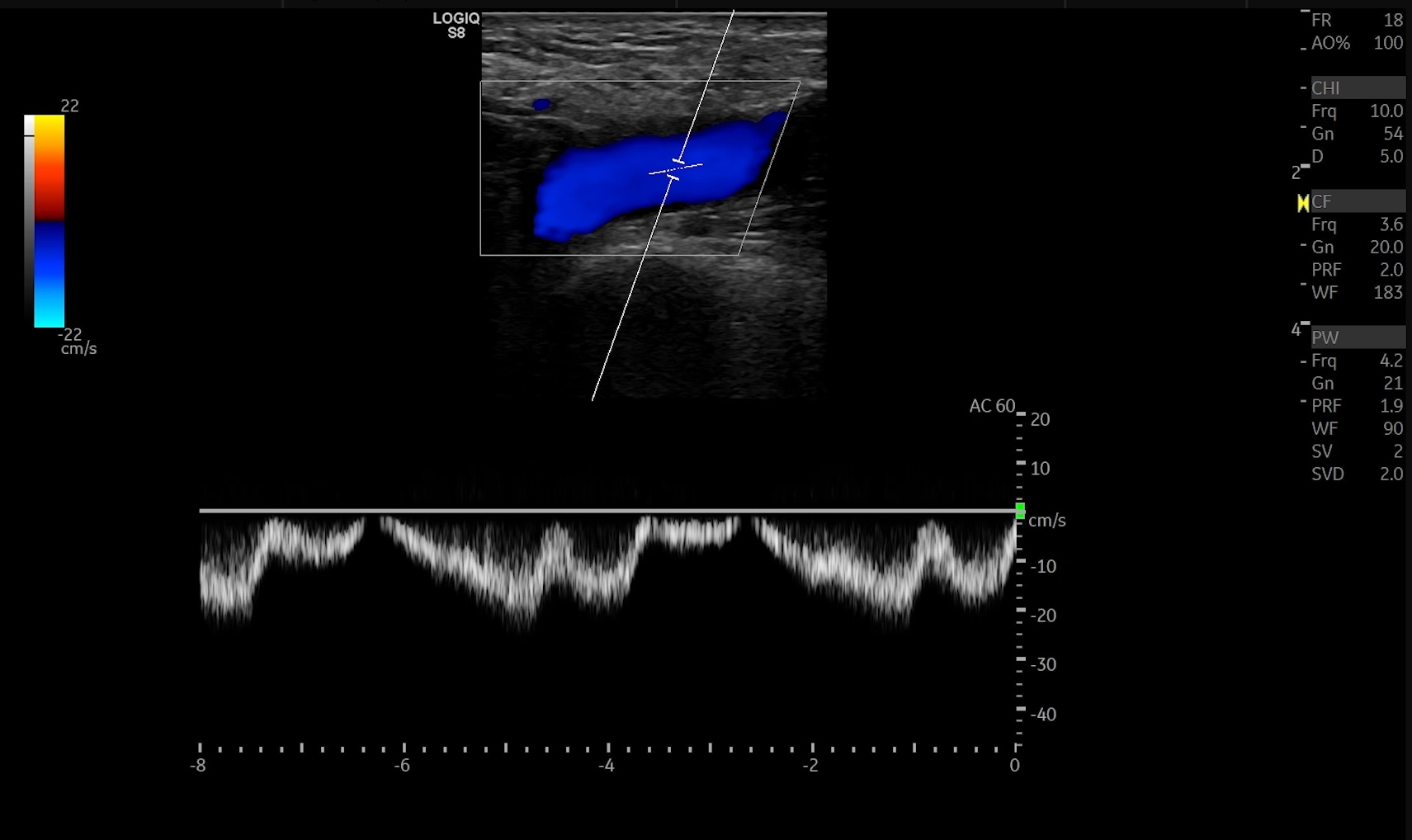
4. Right Common Femoral Vein:
Pulsed-wave Doppler, longitudinal view, measure peak velocity to assess for flow variation
Longitudinal View
5. Left Common Femoral Vein:
B-mode, diameter measurements from anterior to posterior wall and medial to lateral wall at the largest diameter
Transverse View
6. Left Common Femoral Vein:
B-mode, diameter measurements from anterior to posterior wall at the largest diameter
Longitudinal View
7. Left Common Femoral Vein:
Color Doppler
Longitudinal View
8. Left Common Femoral Vein:
Pulsed-wave Doppler, longitudinal view, measure peak velocity to assess for flow variation
Longitudinal View